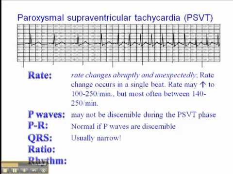
Patients present with syncopal episodes ventricular tachycardia including torsade de pointes ventricular fibrillation and sudden cardiac arrest. The onset of the SVT and offset of the SVT run.
Supraventricular Tachycardia Svt Litfl Ecg Library Diagnosis
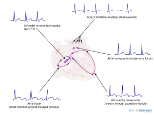
What most people really mean when they call a rhythm SVT is AV Nodal Reentrant Tachycardia or AVNRT which is a reentrant rhythm in or around the AV node.

Svt ecg meaning. Premature beats may be found in healthy individuals as well as patients with underlying heart disease. Unfortunately the electrocardiographic differentiation of VT from SVT with aberrancy is not always possible. If you manage to have the test done during an attack of SVT the ECG.
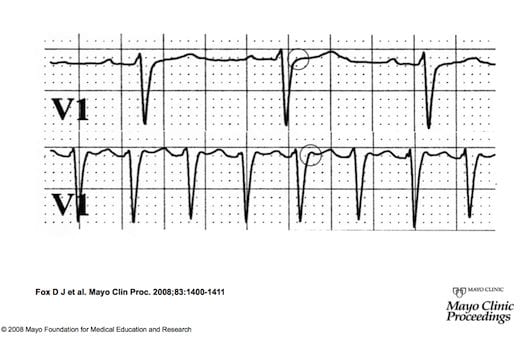
It takes about five minutes and is painless. P wave if sinus or atrial origin no P wave if nodal origin or changes in the P wave such as. ECG Library Homepage Regular broad complex tachycardias can be ventricular VT or supraventricular SVT with aberrancy in origin and differentiation between the two will significantly influence your management of the patient.
ECGs are usually done in hospital or in your GPs surgery. How thats defined may depend on your age and physical condition. With SVT the heart beats more than 100 times a.
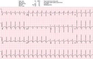
Example of Supraventricular Tachycardia SVT As an ECG technician it is recommended that you capture. ECG changes are stable over time and accentuated during exercise. The term supraventricular tachycardia SVT refers to any tachydysrhythmia arising from above the level of the Bundle of His and encompasses regular atrial irregular atrial and regular atrioventricular tachycardias It is often used synonymously with AV nodal re-entry tachycardia AVNRT a form of SVT In the absence of aberrant conduction eg.
When WPW becomes tachycardic the patient may start to become symptomatic. Irregularity remains Atrial Flutter. Your heart rate during SVT may be as high as 250 beats per minute but is usually between 140 and 180.
The ECG diagnosis of VT is generally straightforward but it does require that this condition be distinguished from aberrantly conducted supraventricular tachycardia SVT which has a similar ECG. Generally speaking for adults a heart rate of more than 100 beats per minute BPM is considered too fast. ECG Library Homepage Regular broad complex tachycardias can be ventricular VT or supraventricular SVT with aberrancy in origin and differentiation between the two will significantly influence your management of the patient.
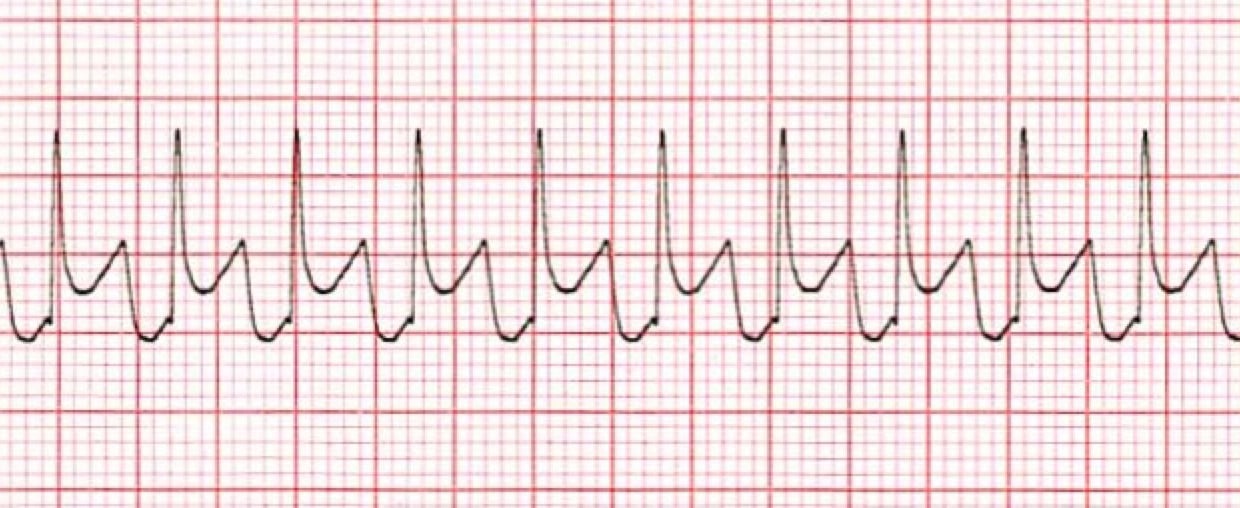
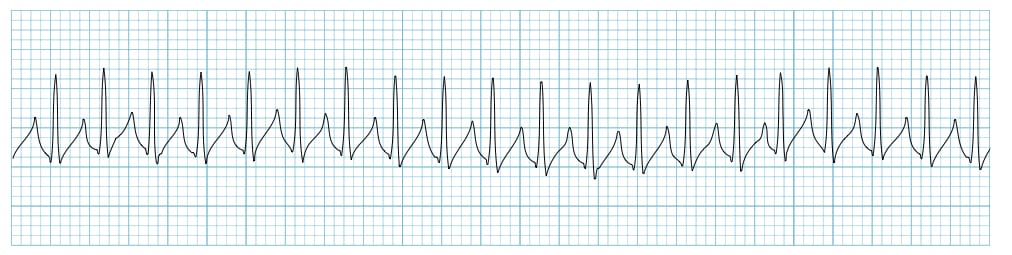
ECG strip showing tachycardia. Supraventricular Tachycardia SVT generally indicates a pathological disease in the Atria or AV Node that can result in severe symptoms to the patient. Supraventricular tachycardia SVT are a group of tachycardias in which at least a structure above the bundle of His is necessary for its continuance.
ECG interpretation must as always proceed systematically in order. The heart normally beats 60 to 100 beats per minute. Atria SVT absent slows down rate.
Regular sometimes alternating block 250-350 bpm 75-150 bpm 31 or 21 block is most common atria SVT negative sawtooth in lead II temporary reduced conduction eg. The ECG is invaluable in the setting of tachycardia. Regular 180-250 bpm 180-250 bpm AV-node SVT in QRS complex R stops Atrial Tachycardia.
Although it is often difficult to arrive at a definitive diagnosis the ECG will allow for a more or less certain diagnosis in most cases. Tachycardia refers to a heart rate thats too fast. Runs with fastest rate and longest duration.
Supraventricular Ectopic Beat SVE A beat that is premature narrow in width but may be slightly different shaped than the patients normal beats. View an animation of tachycardia. ECG strip showing a normal heartbeat.
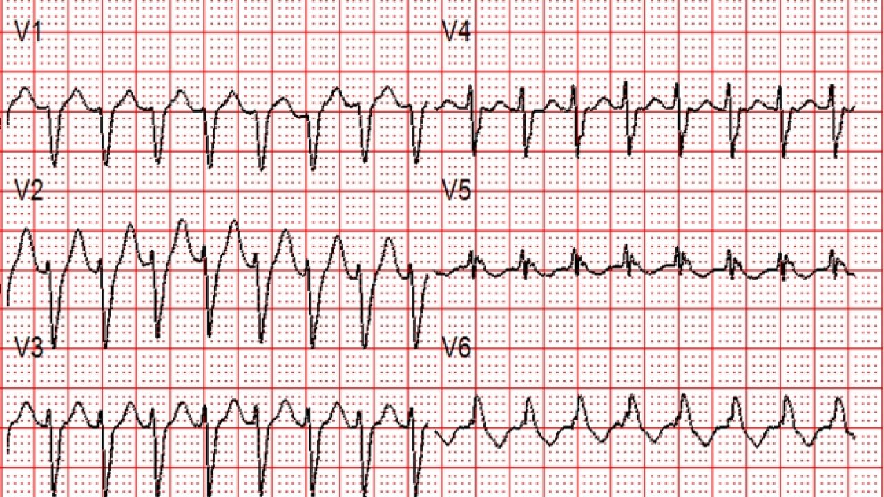
The ECG is characterized by deep and persistent concave-upward ST-segment depression in multiple limb and chest leads. These beats may be sinus atrial or nodal in origin. Bundle branch block the ECG will.
Supraventricular tachycardia SVT is a type of abnormally fast heartbeat. Supraventricular tachycardias include a number of disorders atrioventricular nodal reentrant tachycardia and atrioventricular reentry tachycardia being the most frequent. The heart normally beats 60 to 100 beats per minute while you are at rest and awake.
We also include in this article atrial tachycardia and Inappropriate. However in WPW as discussed earlier there is an additional pathway that skips over the AV node creating a slope in the ECGEKG. Signs and Symptoms as well as treatment of WPW are the same as all PSVTs AKA SVT and so I will cover them as just SVT.
This arrhythmia is usually stable and the prognosis is much more favorable than VT. Supraventricular premature beats are atrial contractions triggered by ectopic foci rather than the sinoatrial nodeThey arise within the atria atrial premature beats or through retrograde conduction in the atrioventricular node junctional premature beats.

Em Cases Ecg Cases 19 Tachycardias

What Is Supraventricular Tachycardia Svt Svt Heart Ventricular Tachycardia Cardiac Nursing

Automatic Tachycardia Wikipedia
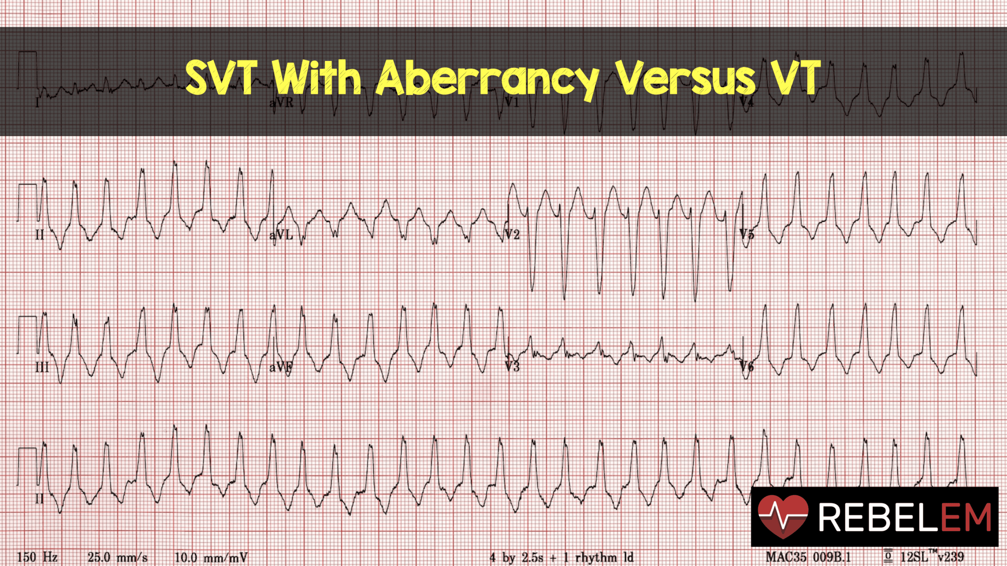
Svt With Aberrancy Versus Vt Rebel Em Emergency Medicine Blog

Supraventricular Tachycardia Svt Litfl Ecg Library Diagnosis

Ekg Atrial Flutter Rush Emergency Medicine
Supraventricular Tachycardia Svt Litfl Ecg Library Diagnosis
Supraventricular Rhythms Ecgpedia
Em Cases Ecg Cases 19 Tachycardias
Ecg Paroxysmal Supraventricular Tachycardia Psvt Youtube

Ekg Ecg Svt Vs Atrial Fibrillation The Ekg Guy Www Ekg Md Youtube
Supraventricular Tachycardia Training Cardiac Arrhythmias Video Propals
Supraventricular Tachycardia Svt Acls Algorithms Com
Supraventricular Tachycardia Vs Sinus Tachycardia Epomedicine
Svt With Aberrancy Or Ventricular Tachycardia Acls Medical Training
Supraventricular Tachycardia Svt Litfl Ecg Library Diagnosis
Supraventricular Tachycardia Svt Litfl Ecg Library Diagnosis

Supraventricular Tachycardia Svt Litfl Ecg Library Diagnosis

Supraventricular Tachycardia Svt Acls Algorithms Com
