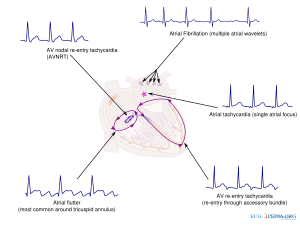
Supraventricular tachycardia SVT is a type of abnormal heart rhythm that results in fast heartbeats. Atrial fibrillation paroxysmal supraventricular tachycardia PSVT atrial flutter and WolffParkinsonWhite syndrome.
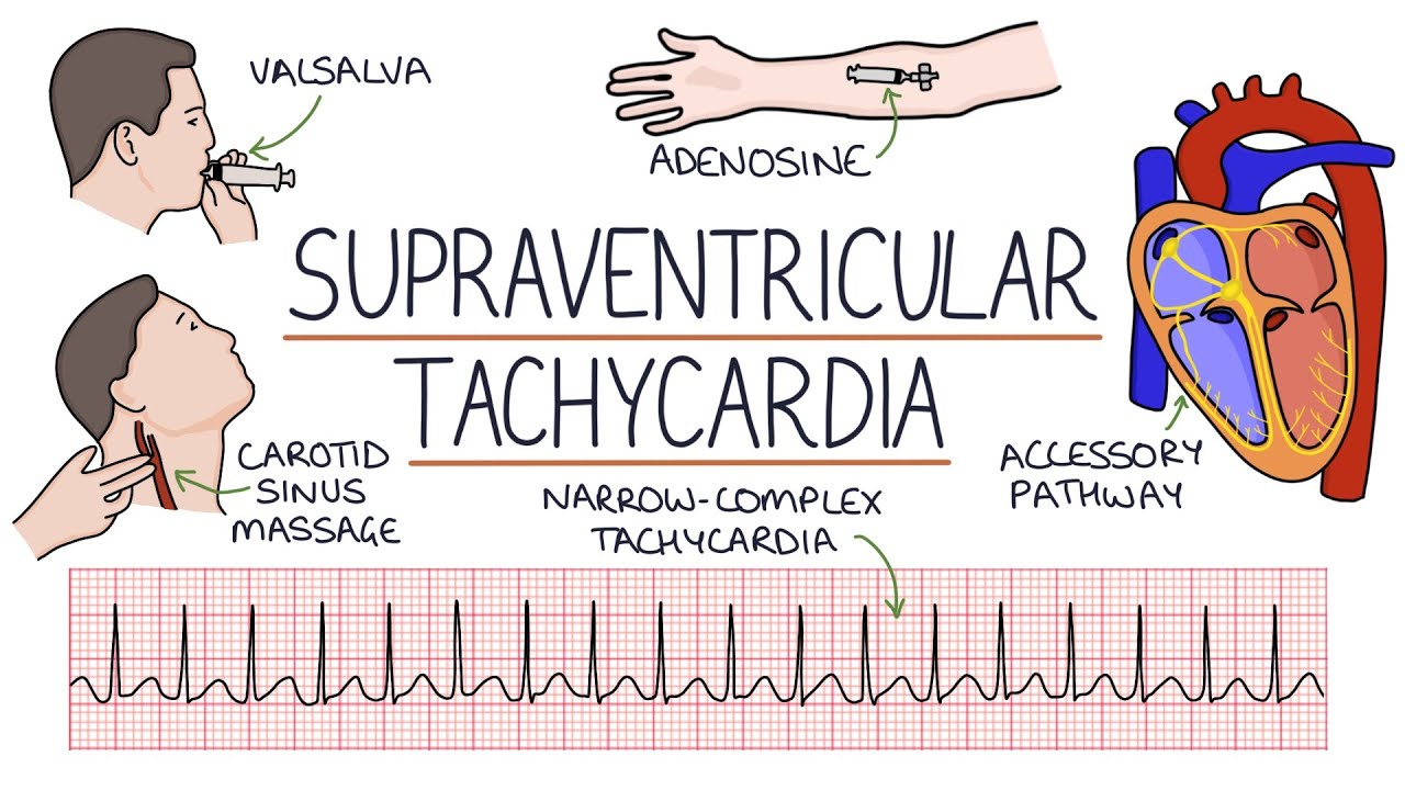
Understanding Supraventricular Tachycardia Svt Youtube
A wide range of conditions may cause ventricular tachycardia and the ECG is as nuanced as are those conditions.
Svt ecg explained. What is supraventricular tachycardia SVT. The term supraventricular tachycardia SVT refers to any tachydysrhythmia arising from above the level of the Bundle of His and encompasses regular atrial irregular atrial and regular atrioventricular tachycardias. We also include in this article atrial tachycardia and Inappropriate.
There are four main types. Supraventricular tachycardias include a number of disorders atrioventricular nodal reentrant tachycardia and atrioventricular reentry tachycardia being the most frequent. Ventricular tachycardia refers to a wide QRS complex heart rhythm that is a QRS duration beyond 120.
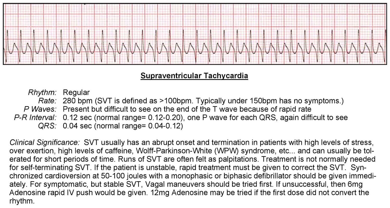
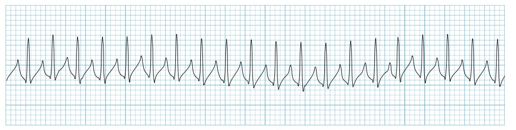
What most people really mean when they call a rhythm SVT is AV Nodal Reentrant Tachycardia or AVNRT which is a reentrant rhythm in or. By definition supraventricular tachycardia must be fast. P waves represent atrial depolarisation.
The PR interval begins at the start of the P wave and ends at the beginning of the Q wave. With SVT the heart beats more than 100 times a minute. Supraventricular tachycardia SVT is a fast heart rhythm arising from abnormal electrical activity in the upper part of the heart.
Supraventricular tachycardia SVT are a group of tachycardias in which at least a structure above the bundle of His is necessary for its continuance. In many cases the underlying mechanism can be deduced from electrocardiography during tachycardia comparing it with. Ventricular tachycardia is a highly nuanced arrhythmia which originates in the ventricles.
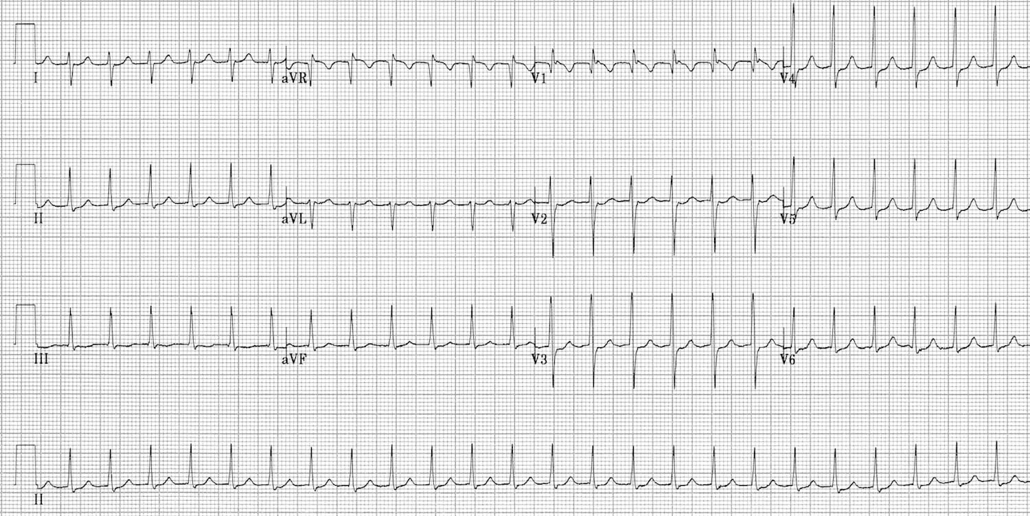
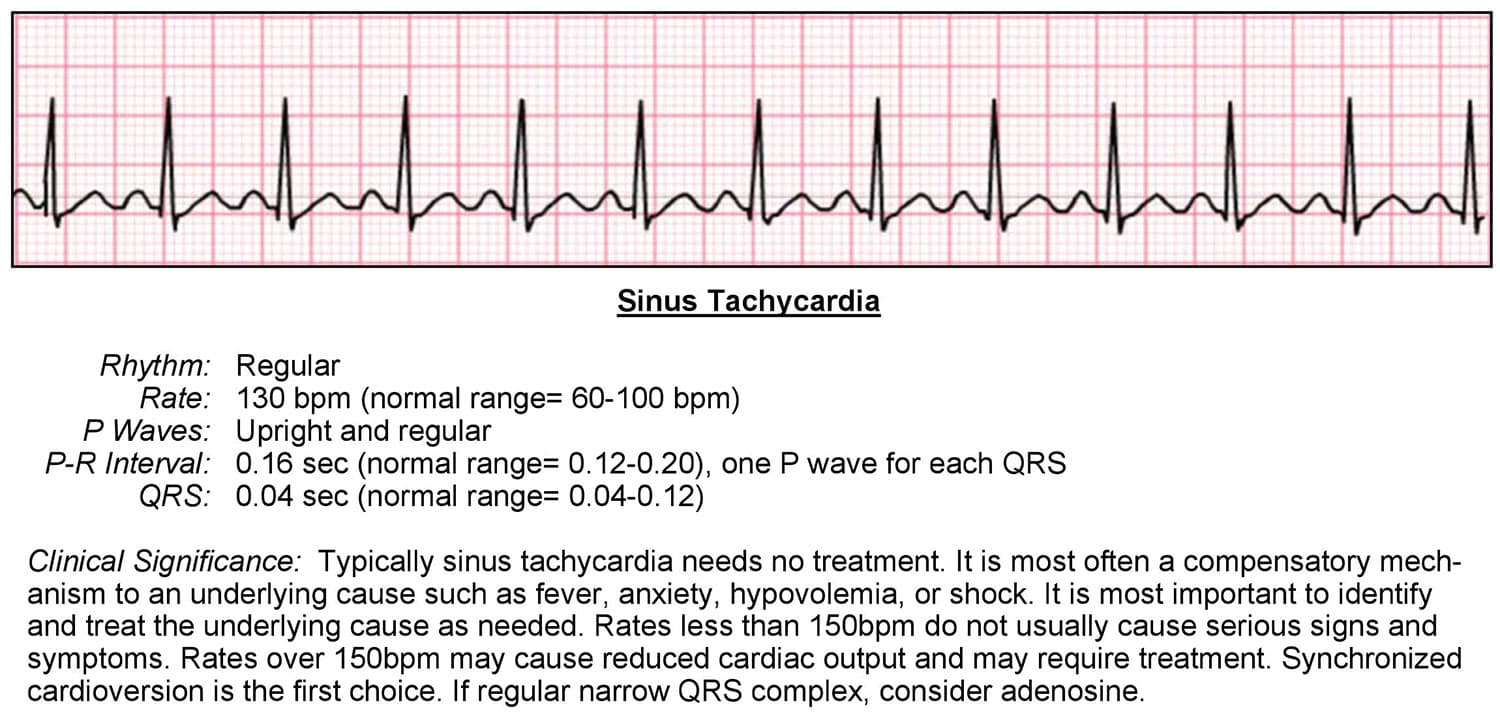
This is the ecg that shows up on exams and the ecg that the triage nurse generally recognizes and leads to the patient being whisked off to the resuscitation bay. The heart normally beats 60 to 100 beats per minute while you are at rest and awake. This is the SVT that we all know and love.
In reality sinus tachycardia is a form of SVT and the rate can easily exceed 150. Supraventricular tachycardia SVT is a category of rapid cardiac arrhythmias that originate in the atrial chambers of the heart. This rhythm is usually narrow since it originates above the ventricles.
An abnormally fast heart rhythm tachycardia can arise from the upper or lower chambers of the heart or be a circuit. The electrical signal passes to the bottom of the heart through a special junction called the AV node. Symptoms common to them all may include palpitations feeling of.
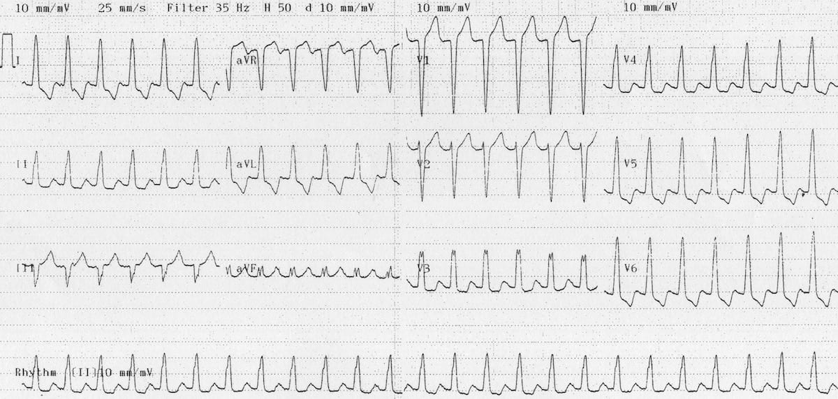
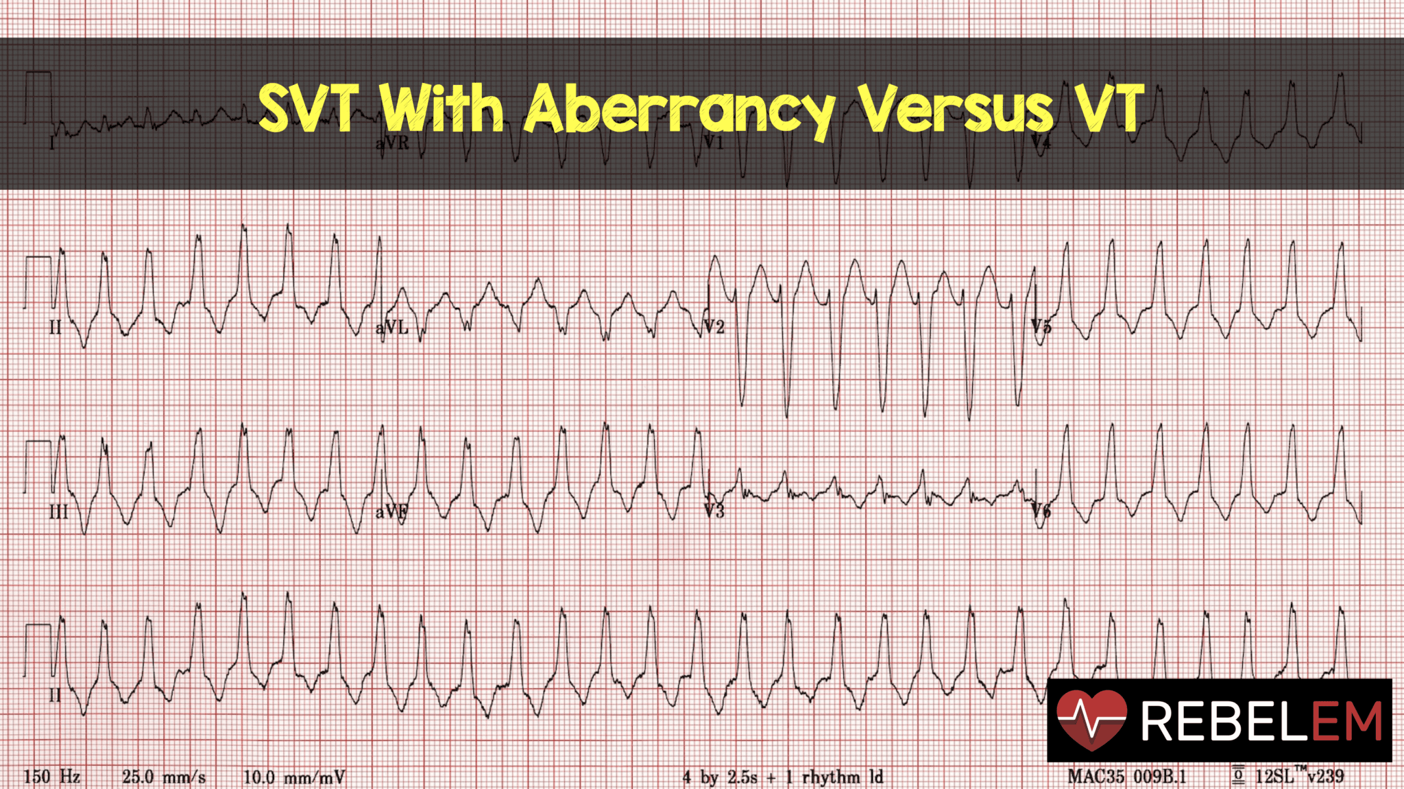
Although there is a broad complex tachycardia HR 100 QRS 120 the appearance in V1 is more suggestive of SVT with aberrancy given that the the complexes are not that broad 160 ms and the right rabbit ear is taller than the left. Studying for a nursing school exam. It may even beat over 200 times a minute.
However on closer inspection there are signs of AV dissociation with superimposed P waves visible. This ECG is a difficult one. Regardless of etiology and ECG ventricular tachycardia is always a potentially life-threatening arrhythmia which requires immediate attention.
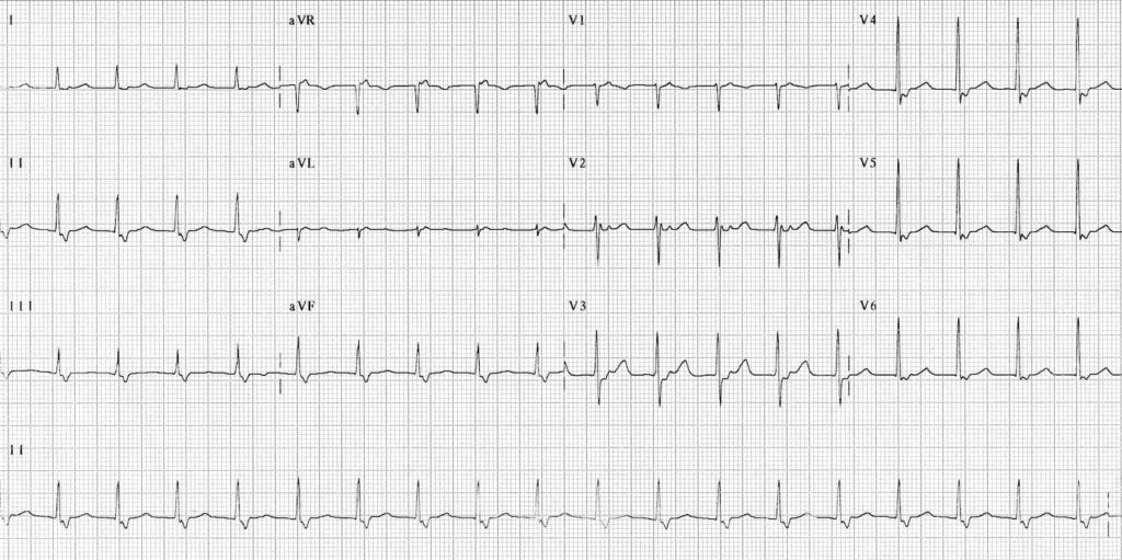
What is Supraventricular Tachycardia SVT. The most common SVTs include atrioventricular nodal re-entrant tachycardia atrioventricular re-entrant tachycardia and atrial tachycardia. In healthy individuals there should be a P wave preceding each QRS complex.
Supraventricular tachycardia SVT is a common cause of hospital admissions and can cause significant patient discomfort and distress. Ventricular Tachycardia VT ECG Review. Supraventricular simply means above the ventricles There are several different kinds of SVT but to a person who has SVT all of.
There are possibly six subdivisions of PSVT. A good rule of thumb to estimate the maximum sinus rate is 220 minus age but that can vary by 10-15 which is a lot. In the absence of aberrant conduction.
Parts of the ECG explained P waves. Click the link below to get FREE access to a massive library of helpful video. A normal heartbeat originates from the sinus node the hearts pacemaker.
It is often used synonymously with AV nodal re-entry tachycardia AVNRT a form of SVT. It represents the time taken for electrical activity to move between the atria and the ventricles.
Vt Versus Svt Litfl Medical Blog Ecg Library Basics
Tachycardia Acls Wiki

Svt On The Ecg Usf Emergency Medicine Residency

Supraventricular Tachycardia Svt Acls Algorithms Com
Supraventricular Tachycardia Svt Litfl Ecg Library Diagnosis

44 Paroxysmal Supraventricular Tachycardia Ekg Interpretation Nursing Students School Health

What Is Supraventricular Tachycardia Svt Svt Heart Ventricular Tachycardia Cardiac Nursing

Ekg Ecg Svt Vs Atrial Fibrillation The Ekg Guy Www Ekg Md Youtube
Supraventricular Tachycardia Svt Litfl Ecg Library Diagnosis

Svt On The Ecg Usf Emergency Medicine Residency
Svt With Aberrancy Versus Vt Rebel Em Emergency Medicine Blog

Supraventricular Tachycardia Svt Litfl Ecg Library Diagnosis
Tachycardia Acls Wiki
Supraventricular Tachycardia Svt Litfl Ecg Library Diagnosis
Supraventricular Rhythms Ecgpedia

Svt On The Ecg Usf Emergency Medicine Residency
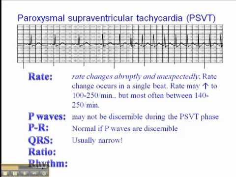
Ecg Paroxysmal Supraventricular Tachycardia Psvt Youtube

Supraventricular Tachycardia Svt Litfl Ecg Library Diagnosis

Vt Ventricular Tachycardia Vs Svt Supraventricular Tachycardia With Aberrancy Youtube
